Research at

Ailles Lab

Get Started

Welcome to Ailles Lab

Dr. Laurie Ailles and her team are focusing on three major research areas:

  • Identification of prognostic biomarkers and novel targets for head and neck squamous cell carcinoma (HNSCC) patients
  • Epigenetics in clear cell renal cell carcinoma (ccRCC)
  • Targeting cancer-associated fibroblasts (CAFs) in solid tumors
See More

What We Do

Dr. Laurie Ailles and her team are focusing on three major research areas:

  • Identification of prognostic biomarkers and novel targets for head and neck squamous cell carcinoma (HNSCC) patients
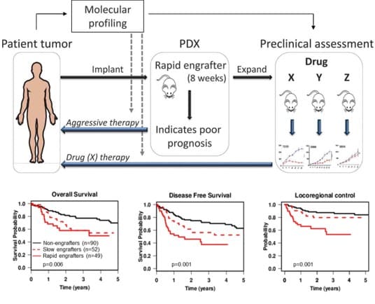
  • Individualized treatment decisions are difficult in HNSCC due to the lack of prognostic and predictive biomarkers. Poor HNSCC survival rates clearly indicate a need for new and better therapies. Dr. Ailles and her team are working to identify prognostic biomarkers for patient stratification in HNSCC using genomic characterization of primary tumor tissues in combination with their functional behavior in tumor engraftment assays in immune-compromised mice. They are focused on identifying novel targets, pathways, and candidate drugs for the treatment of the most aggressive HNSCC tumors using patient-derived xenografts.

  • Epigenetics in clear cell renal cell carcinoma (ccRCC)
  • The Ailles team uses primary patient-derived cultures and xenografts to study the epigenetic deregulation occurring in ccRCC. More specifically, they focus on identifying the downstream effects on target genes and pathways, and identifying small molecules with rapid clinical translational potential.

  • Targeting cancer-associated fibroblasts (CAFs) in solid tumors
  • Cancer-associated fibroblasts (CAFs) are known to play a role in promoting cancer proliferation, invasion, and chemo-resistance through acquisition of an “activated” state. The Ailles team has developed methods for the isolation of CAFs from primary patient-derived tumors and carried out molecular characterization of these tumours to identify molecular interactions between CAFs and cancer cells that may represent therapeutic targets. They have demonstrated functional heterogeneity within the CAF population, and are continuing to investigate strategies for epigenetic reprogramming of CAFs to a less activated state.

For a complete list of publications, click here: https://www.ncbi.nlm.nih.gov/pubmed?term=ailles+l[au]

Research Overview

Icon image of MRI

Utilizing Patient-Derived Xenografts to Identify Prognostic Biomarkers and Novel Therapies for Oral Squamous Cell Carcinoma (OSCC) Patients

We have shown that the ability of OSCC tumor tissues to initiate engraftment in immune-compromised mice is indicative of worse outcomes in the corresponding patients (Karamboulas et al, Cell Reports, 2018). Even stronger prognostic potential is obtained when the engraftment kinetics are monitored, with “rapid engrafters” having particularly poor outcomes. We have also carried out treatment of patient-derived xenograft (PDX) models with targeted therapies, allowing us to identify responsive and non-responsive models, and to correlate responses with genetic profiling (Karamboulas et al, Cell Reports, 2018; Ruicci et al, Int J Cancer, 2019). We are now building on these results in the following ways:

  • Validation of rapid engraftment as a functional prognostic biomarker in a prospective study
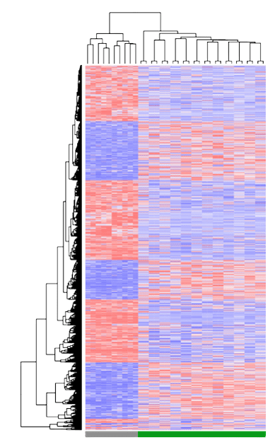
  • Genetic and proteomic profiling of rapid engrafters vs non-engrafters to identify prognostic biomarkers
  • Pharmacogenomic analysis of rapid engrafters to identify novel candidate therapies
  • Interrogation of therapy responses (both standard-of-care and novel candidates) in existing PDX models
  • Development of predictive biomarkers (for both standard-of-care and novel therapies)

Characterizing the Role of Carcinoma-Associated Fibroblasts in Head and Neck Squamous Cell Carcinoma (HNSCC) Invasion

Cancer-associated fibroblasts (CAFs) are an important component of the tumor microenvironment and play a multi-faceted role in tumor progression. CAFs have been shown to promote many of the “hallmarks of cancer”, including invasion and metastasis. We have developed methods in the lab to isolate CAFs from primary tumor tissues and adjacent normal tissues of HNSCC patients. We have developed a 3-D co-culture spheroid invasion assay that allows us to observe and quantify CAF-induced invasion of HNSCC cells. We are currently carrying out molecular profiling of CAFs and cancer cells upon co-culture to identify targetable key molecular interacting partners, and investigate their role in invasion using our 3-D invasion assay.

CAFs represent fibroblasts that have been “activated” via an epigenetic switch. Thus an alternative approach to targeting CAFs is to target their epigenetic state. We are currently working with the Structural Genomics Consortium (SGC; http://www.thesgc.org) to screen an epigenetic chemical probe library for the ability to inhibit or reduce the activation-associated properties of CAFs.

Icon image of MRI
Icon image of MRI

Functional and Molecular Characterization of Cancer-Associated Fibroblasts in High Grade Serous Ovarian Cancer (HGSOC)

To enable molecular analysis of CAFs isolated directly from tumor samples, we recently identified CD49e as a definitive HGSOC CAF marker. Transcriptional profiling of isolated CD49e+ CAFs revealed that patients stratify into two groups based on their CAF gene signatures: One with high Fibroblast Activation Protein expression(FAP-High; FH) and one with low FAP expression (FAP-Low; FL). Immunofluorescence and flow cytometry revealed that HGSC patients contain a mixture of FH and FL CAFs at varying ratios. RNA-seq on purified FH and FL CAFs was used to generate gene signatures that could identify FH and FL patients within The Cancer Genome Atlas RNAseq data set. FH patients had significantly shorter disease-free and overall survival than FL patients. Similarly, patients from our own Institute with predominantly FH CAFs relapsed significantly more rapidly than patients with predominantly FL CAFs. A comprehensive array of functional assays on FH and FL CAFs showed that FH (but not FL) CAFs promoted chemo-resistance and invasion of cancer cells in culture, and promoted tumor growth and metastasis in mice ( https://doi.org/10.1101/519728). In addition, we have identified a transcription factor, TCF21, which suppresses the FH state. Our current projects are focused on further understanding the role of TCF21 in regulating the CAF state in HGSOC, as well as gaining further insights into the functional roles of distinct CAF subtypes in this cancer.

The Role of Protein Arginine Methyltransferase 1 (PRMT1) in Clear Cell Renal Cell Carcinoma

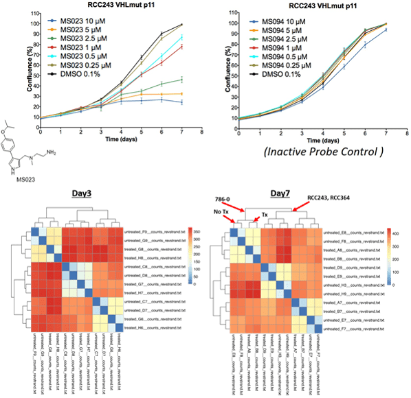
Sporadic Renal Cell Carcinoma (RCC) is dominated by the clear cell subtype (ccRCC) and overwhelmingly associated with a biallelic inactivation of the von Hippel-Lindau (VHL) gene leading to constitutive activation of the cell's hypoxia response and deleterious alterations to gene expression, metabolism and growth characteristics. However, VHL inactivation alone is insufficient to cause tumourigenesis. Other key genetic players identified through sequencing of ccRCC patient cohorts include frequent inactivating mutations in epigenetic regulatory enzymes - proteins that mediate the accessibility of transcription machinery to specific areas of the genome. The high frequency of these alterations in ccRCC implicate epigenetic vulnerabilities that may be exploited to develop new therapies. The Ailles Lab has generated a panel of patient-derived cell lines from ccRCC patients (Lobo et al, BMC Cancer, 2016). In collaboration with the Structural Genomics Consortium (SGC; http://www.thesgc.org) we carried out a screen of an epigenetic chemical probe library for the ability to inhibit the proliferation of our patient-derived ccRCC cell lines. From this screen we identified the compound MS023 as a candidate hit. MS023 has potent activity against the type I protein arginine methytransferase family (PRMT1, 3, 4, 6 and 8). PRMTs transfer methyl groups to both nuclear histones and cytoplasmic targets, influencing gene expression, cell signaling, growth and viability. Specific PRMT3, 4 and 6 inhibitors failed to inhibit ccRCC cell growth in our screen, and PRMT8 is not expressed in this cell type, thus PRMT1 is the primary target for growth inhibition of ccRCC by MS023. We have validated the specificity of MS023 for PRMT1 through overexpression rescue experiments, and have also validated the importance of PRMT1 in ccRCC cell proliferation through knock-down and knock-out studies. Current studies are focused on gaining a mechanistic understanding of the role of PRMT1 in ccRCC growth.

Icon image of MRI

Team

Initial of Dr. Laurie Ailles

Dr. Laurie Ailles

Principal Investigator
picture of Dr. Christina Karamboulas

Dr. Christina Karamboulas

Scientific Associate
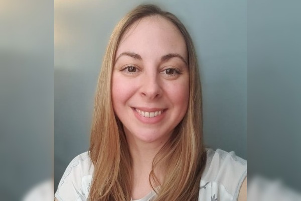
picture of Jalna Meens

Jalna Meens

Lab Manager/ Research Technician
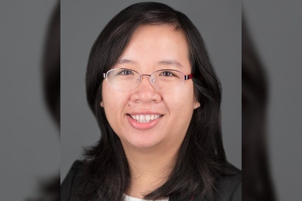
picture of Dr. Ngoc Bui

Dr. Ngoc Bui

Postdoctoral Fellow
picture of Dr. Kunal Karve

Dr. Kunal Karve

Postdoctoral Fellow
picture of Joe Walton

Joe Walton

Ph.D. Candidate
picture of Stephanie Poon

Stephanie Poon

Ph.D. Candidate
picture of Rita Gill

Rita Gill

Research Technician
picture of Karen Arevalo

Karen Arevalo

Ph.D. Candidate
picture of Angel Ng

Angel Ng

Ph.D. Candidate

Recent Publications

Postions

We are not currently recruiting for any new positions in the Ailles Lab.

Resources

Contact Us

Ailles Lab

The Ailles Lab is located at:
101 College Street, Room 12-305
MaRS Building - East Tower
Princess Margaret Cancer Research Tower
Toronto, Ontario, M5G 1L7
Canada

Dr. Laurie Ailles
Principal Investigator
Phone: (+1) 416-581-7868
Email: laurie.ailles@uhn.ca

Lucy Fuccillo
Administrative Assistant
Phone: (+1) 647-882-9298
Email: lucy.fuccillo@uhn.ca

Icons made by Freepik from www.flaticon.com is licensed by CC 3.0 BY